| Shared Root — common ancestor of both branches |
| Root |
1622 |
 Friedrich Leibniz
1597–1652 |
Univ. Leipzig |
Professor of Moral Philosophy; father of Gottfried Leibniz |
|
1643 |
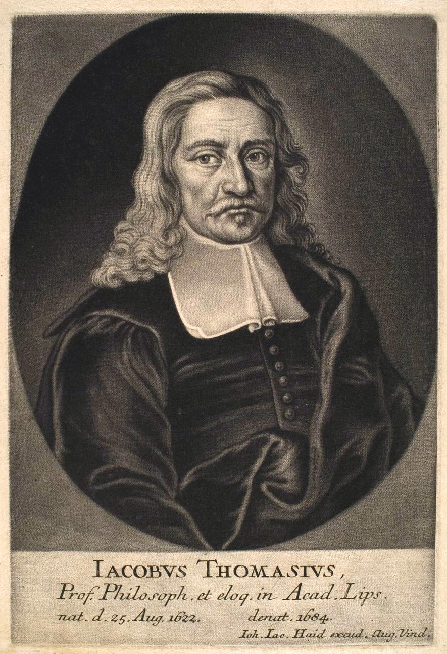 Jakob Thomasius
1622–1684 |
Univ. Leipzig |
Philosopher and jurist; mentor to both Mencke (Gauss line) and Gottfried Leibniz (Euler line) |
| Gauss Branch — via Plücker (1st advisor to Felix Klein) |
| Gauss |
1666 |
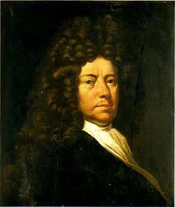 Otto Mencke
1644–1707 |
Univ. Leipzig |
Founded Acta Eruditorum (1682), Germany’s first scientific journal |
|
1685 |
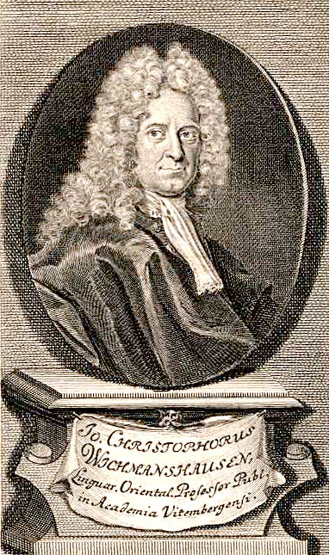 J. C. Wichmannshausen
1663–1727 |
Univ. Leipzig |
Philosopher; dissertation on divorce according to natural law |
|
1713 |
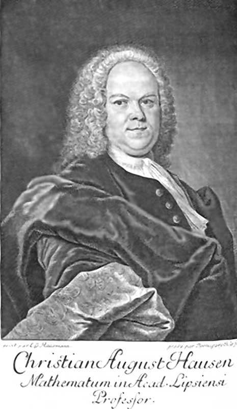 Christian August Hausen
1693–1743 |
Halle-Wittenberg |
Mathematician and physicist; early research on electricity |
|
1739 |
 Abraham G. Kästner
1719–1800 |
Leipzig → Göttingen |
Encyclopedic mathematician; advisor to Pfaff (who advised Gauss) |
|
1786 |
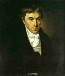 Johann Friedrich Pfaff
1765–1825 |
Göttingen → Helmstedt |
Pfaffian differential systems; Gauss’s formal doctoral advisor |
|
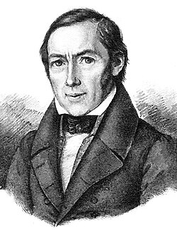
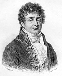
1799 |
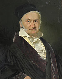 Carl Friedrich Gauss ★
1777–1855 |
Helmstedt → Göttingen |
“Prince of Mathematicians”; fundamental theorem of algebra; 127,000+ descendants |
|
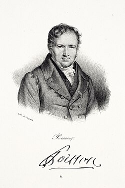
1812 |
 Christian L. Gerling
1788–1864 |
Göttingen → Marburg |
Geodesy and astronomy; extensive correspondence with Gauss |
|
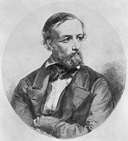
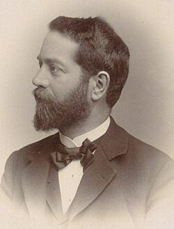
1823 |
 Julius Plücker
1801–1868 |
Marburg → Bonn |
Line geometry; analytic geometry; 1st (primary) advisor to Felix Klein |
| Euler Branch — via Lipschitz (2nd advisor to Felix Klein) |
| Euler |
1666 |
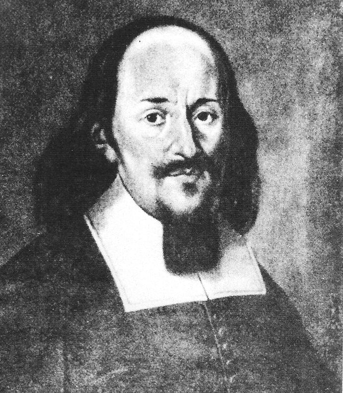
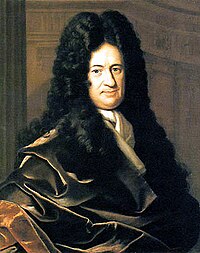 Gottfried W. Leibniz ★
1646–1716 |
Altdorf → Berlin · Hanover |
Co-inventor of calculus; logic; philosophy; 183,000+ academic descendants |
|
~1690 |
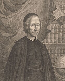 Nicolas Malebranche
1638–1715 |
Paris, Oratory |
French philosopher & mathematician; doctoral advisor of Jacob Bernoulli |
|
1684 |
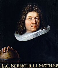 Jacob Bernoulli
1655–1705 |
Univ. Basel |
Bernoulli numbers; probability theory; discovered constant e |
|
1694 |
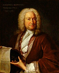 Johann Bernoulli
1667–1748 |
Basel → Groningen |
Calculus of variations; L’Hôpital’s rule; primary teacher of Euler |
|
1726 |
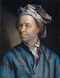 Leonhard Euler ★
1707–1783 |
Basel → St. Petersburg → Berlin |
Most prolific mathematician in history; introduced e, i, π notation |
|
~1754 |
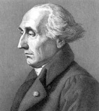 Joseph-Louis Lagrange ★
1736–1813 |
Turin → Berlin → Paris |
Lagrangian mechanics; Lagrange multipliers; number theory |
|
~1800 |
 Jean-Baptiste Fourier ★
1768–1830 |
École Normale, Paris |
Fourier series and transforms; heat equation; co-advisor to Dirichlet |
|
1800 |
 Siméon Denis Poisson ★
1781–1840 |
École Polytechnique |
Poisson distribution; electrostatics; co-advisor to Dirichlet |
|
1827 |
 Gustav Dirichlet ★
1805–1859 |
Bonn → Berlin → Göttingen |
Modern definition of function; Dirichlet series; analytic number theory |
|
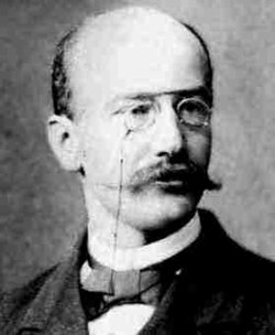
1853 |
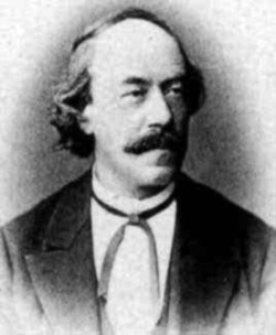 Rudolf Lipschitz
1832–1903 |
Univ. Berlin → Bonn |
Lipschitz continuity; Clifford algebras; 2nd advisor to Felix Klein |
| Both Branches Merge at Felix Klein |
| Merged |
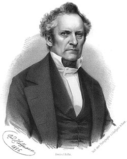
1868 |
 Felix Klein ★
1849–1925 |
Bonn → Erlangen → Göttingen |
Erlangen Program; Klein bottle; 68 PhD students; 87,000+ descendants |
|
1873 |
 Ferdinand von Lindemann ★
1852–1939 |
Erlangen → Königsberg → Munich |
Proved π is transcendental (1882); also advised Hilbert and Minkowski |
|
1891 |
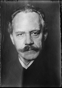 Arnold Sommerfeld ★
1868–1951 |
Königsberg → Munich |
Quantum physics; advised more Nobel Prize winners than any other supervisor |
| Electrical Engineering & Signal Processing Lineage |
| EE/DSP |
1926 |
 Ernst A. Guillemin ★
1898–1970 |
Munich → MIT |
Network analysis and synthesis; IEEE Medal of Honor 1961 |
|
1948 |
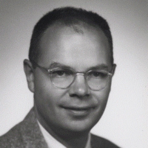 David Fears Tuttle, Jr.
1914–? |
MIT → Stanford |
Network synthesis; authored classic textbooks; Stanford legend |
|
1952 |
 Ernest S. Kuh NAE
1928–2015 |
Stanford → UC Berkeley |
Circuit theory; electronic design automation; Berkeley EECS chair & Dean |
|
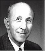
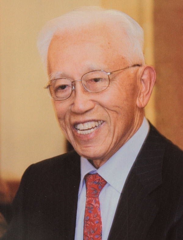
1962 |
 Sanjit K. Mitra NAE
1935–present |
UC Berkeley → UCSB |
Digital signal processing; 12 books, 240+ journal papers; IEEE Education Medal 2006 |
|
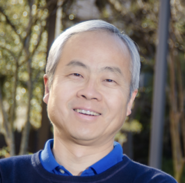
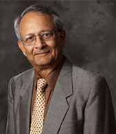
1982 |
 P. P. Vaidyanathan NAE
1954–present |
UCSB → Caltech |
Filter banks and multirate signal processing; IEEE Jack S. Kilby Medal 2024 |
|
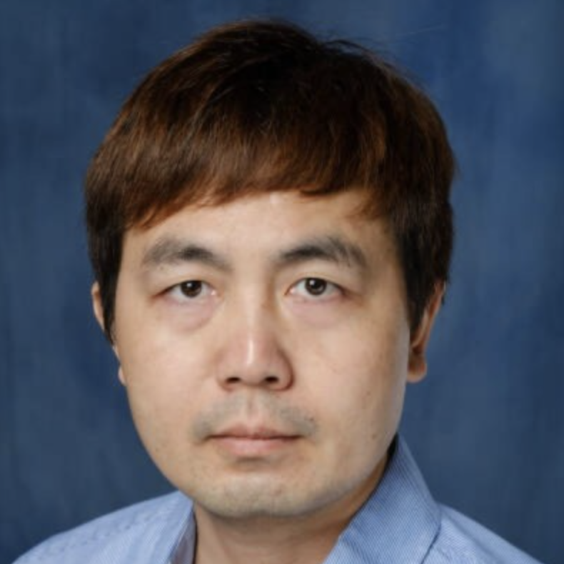
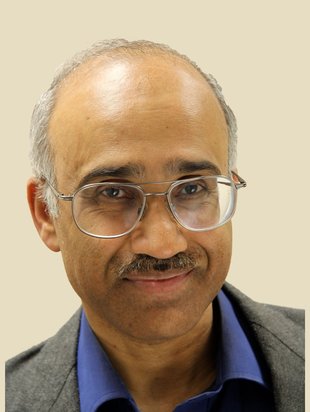
1993 |
 Tsuhan Chen
1966–present |
Caltech → CMU → Cornell → NUS |
Computer vision; Chief Scientist of AI Singapore; ~30 patents |
| Current Generation |
| Present |
2014 |
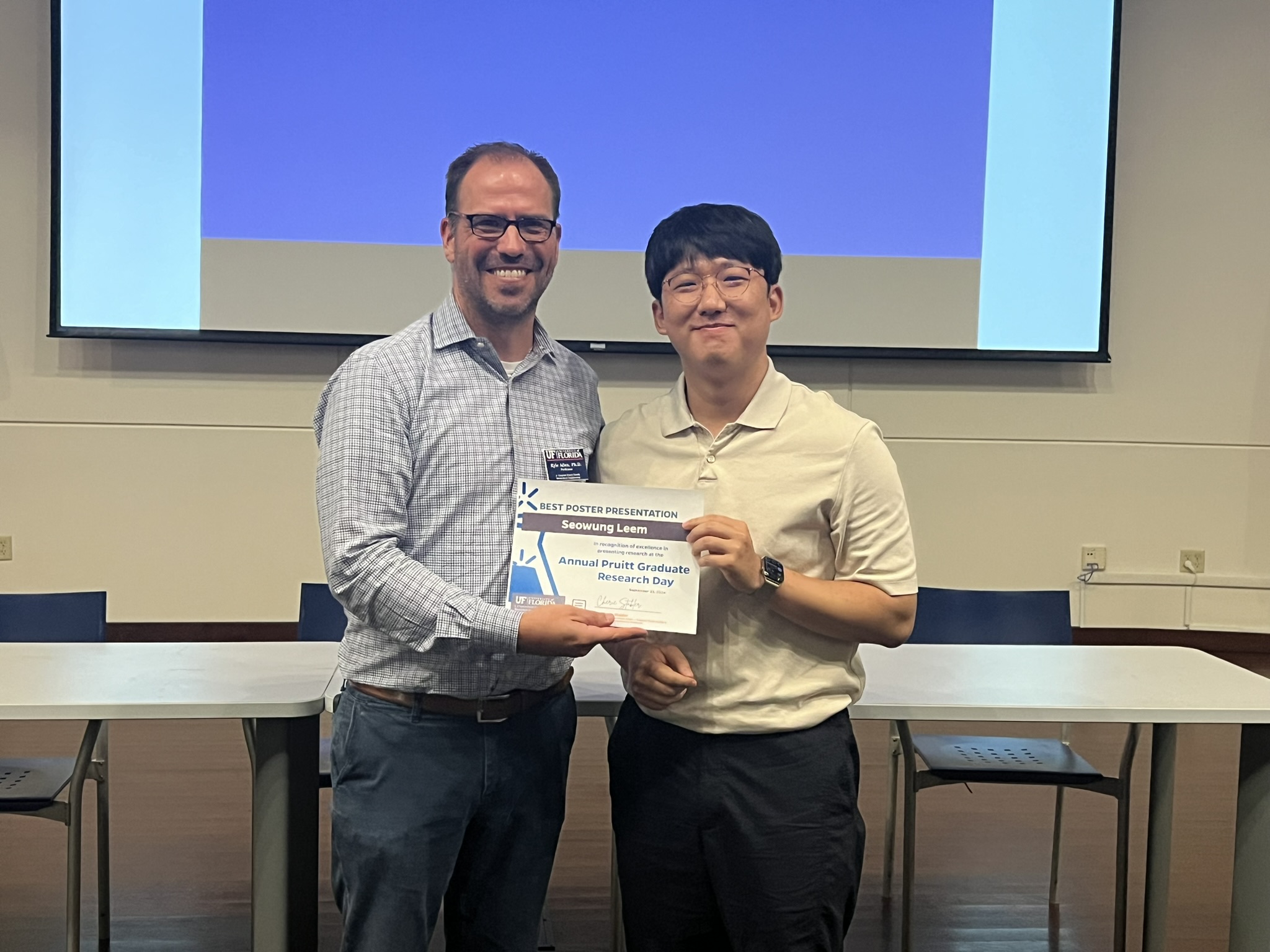
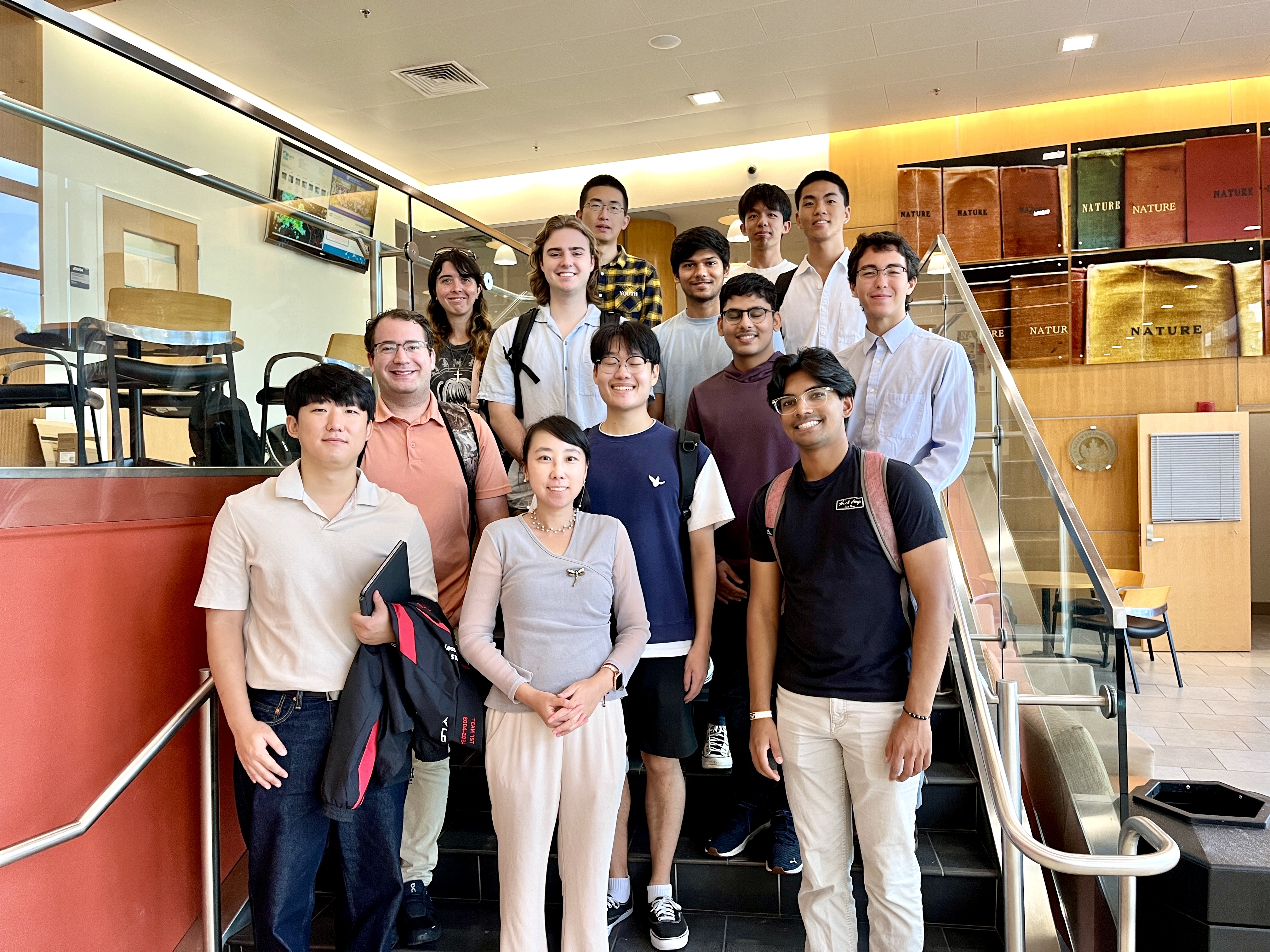
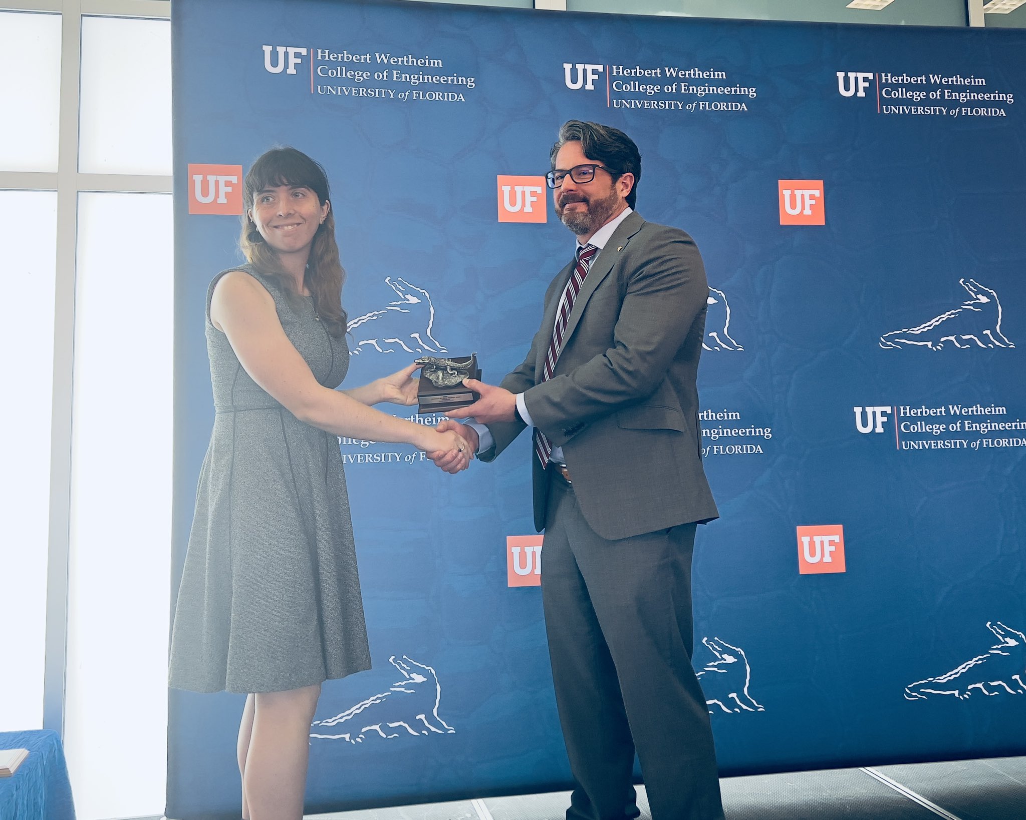
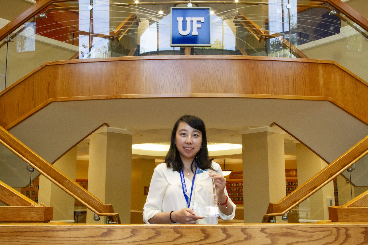
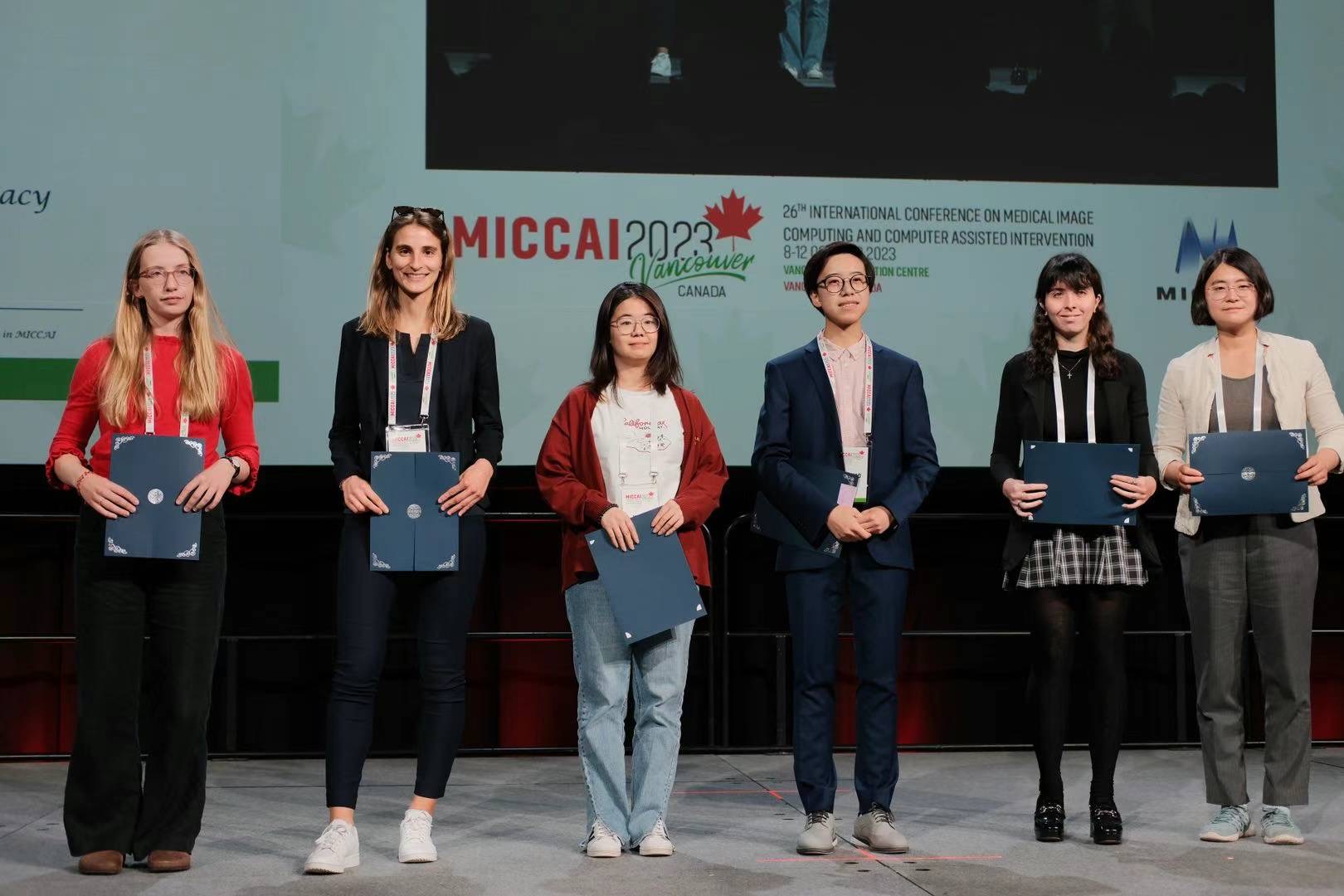
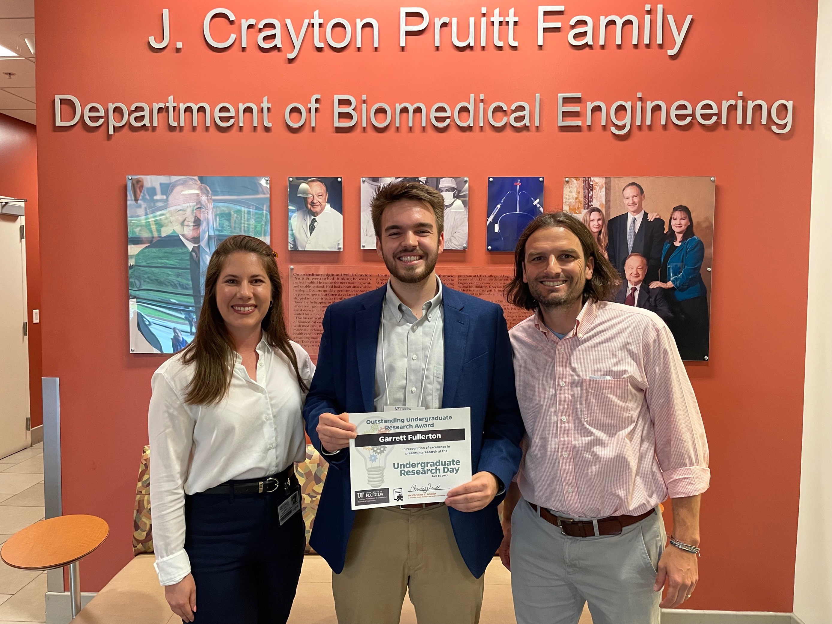
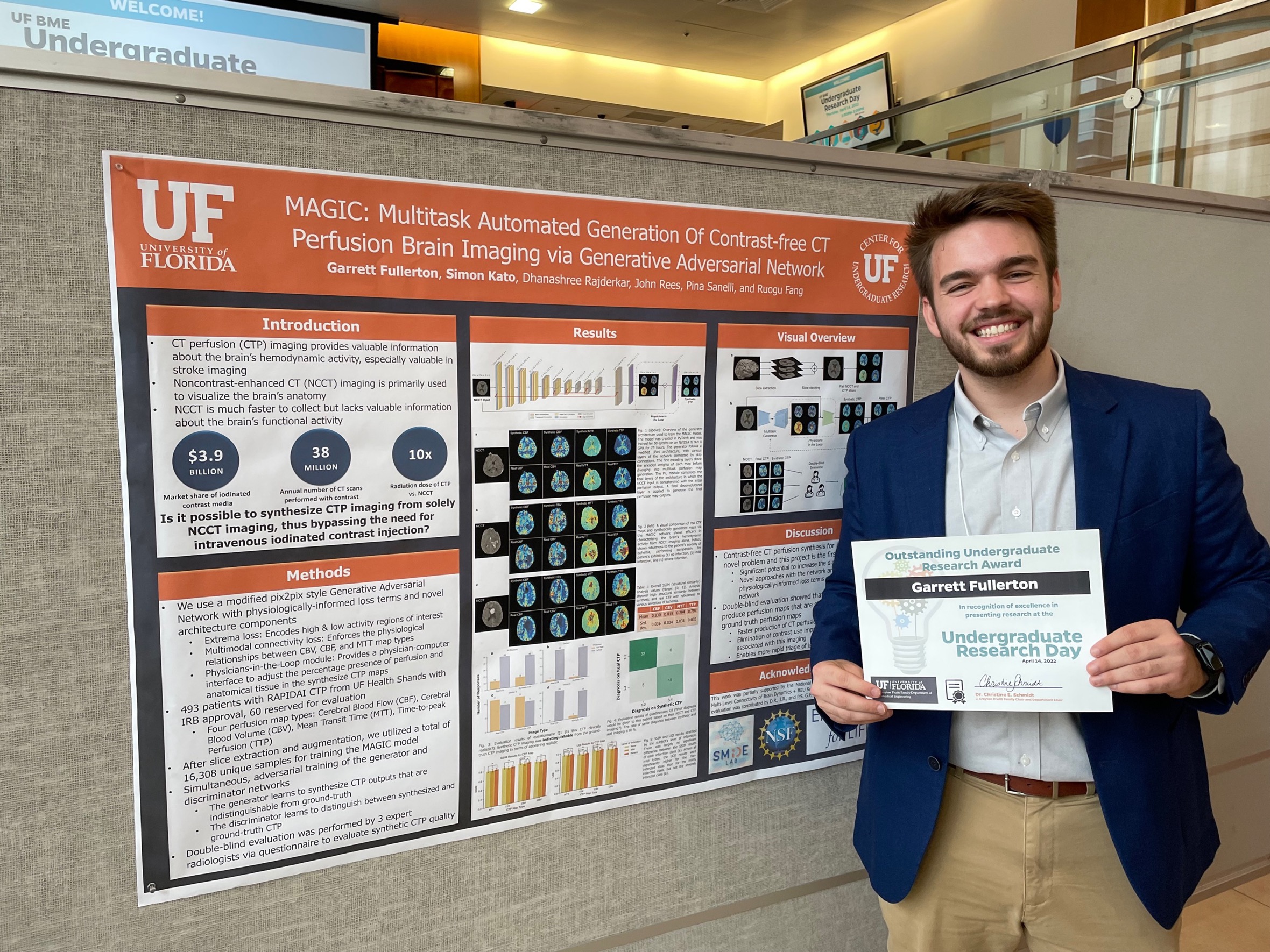
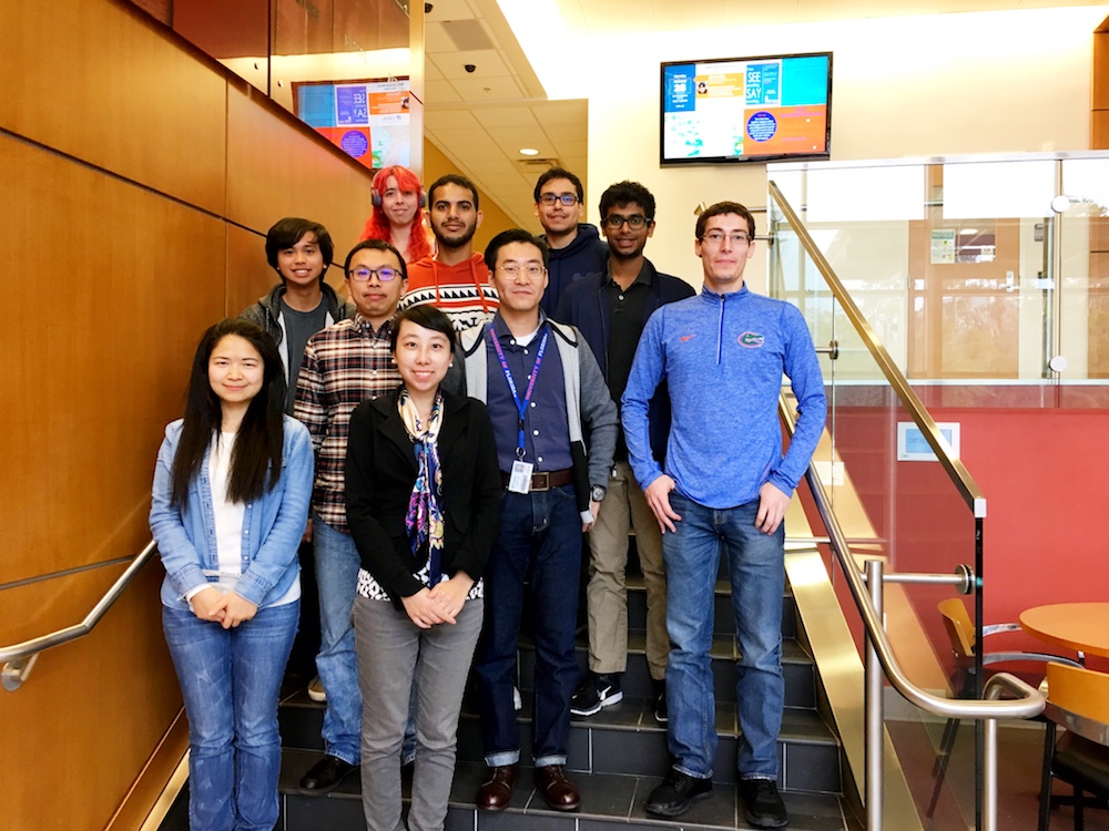
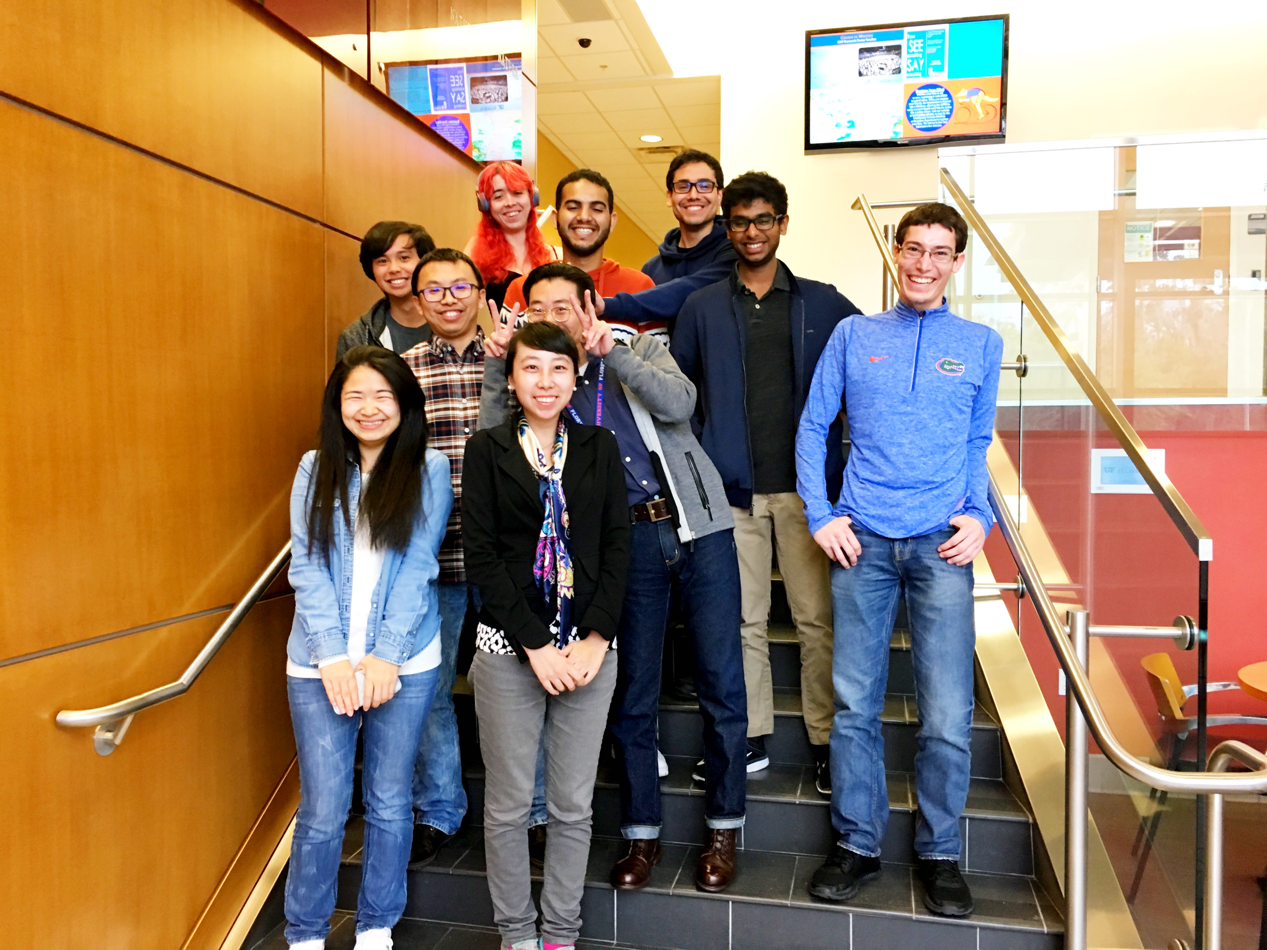
 Ruogu Fang |
Cornell → FIU → Univ. Florida |
Associate Professor, Pruitt Family Endowed Fellow, BME, UF; Director, SMILE Lab |